Chordomas
Chordomas are malignant tumors that develop within the spine or base of the skull. They arise from remnants of embryonic cells that form the spinal column. While they are usually slow growing, they may invade nearby structures or destroy surrounding bone and tissue. Often located next to important arteries and nerves, chordomas can be difficult to treat and tend to return after treatment.

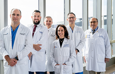
Meet Our Brain Tumor Care Team
Meet the specialists at the Gerald J. Glasser Brain Tumor Center who are experts in treating chordomas.
Meet the TeamSymptoms
As the tumor grows and puts pressure on the surrounding brain or cranial nerves, symptoms may develop or increase in severity, including:
- Weakness or numbness in the limbs or face
- Double vision
- Hearing loss
Chordomas in the spine can cause:
- Tingling, numbness or weakness in the arms or legs
- Loss of bladder or bowel control
Diagnosis
A chordoma is diagnosed through analysis of symptoms, a physical exam and specialized imaging studies of the brain, skull base or spinal column. High-resolution imaging scans with contrast dye are used to help make the tumor visible and identify its size, location and relationship to major arteries. They can also determine the extent of any bone destruction or compression of the brain, cranial nerves or spinal cord.
A needle biopsy may be used to confirm the diagnosis of a spinal column chordoma, often quickly followed by surgery to prevent the spread of any tumor cells.
Treatment
Surgery – Typically, the initial treatment for a chordoma is to remove as much of the tumor as safely possible. Our team will develop a personalized treatment plan based on the size and extent of your tumor and evaluate whether traditional open or minimally invasive endoscopic skull base surgical techniques should be used. Depending on the tumor’s location, the surgical team may include neurosurgeons, otolaryngologists (ENT) and plastic, urologic and vascular surgeons.
Post-surgical treatment – Chordomas tend to return after treatment, so follow-up care is particularly important. Each patient’s case is reviewed by our tumor board after surgery. Together, the multidisciplinary team of experts recommends the best personalized treatment options for you. These incorporate the latest treatment protocols and participation in national clinical trials.
Follow-up treatments may include:
- Observation – Even when the tumor has been totally removed, surveillance MRI or CT scans are used to watch for recurrence.
- Stereotactic radiosurgery (CyberKnife®) – This non-invasive treatment option is used after surgery for small or residual tumors in hard-to-reach locations or if a tumor returns after surgery.
Our team will closely monitor you and personalize your follow-up care. Our patient navigator will also connect you with our support group and other resources.